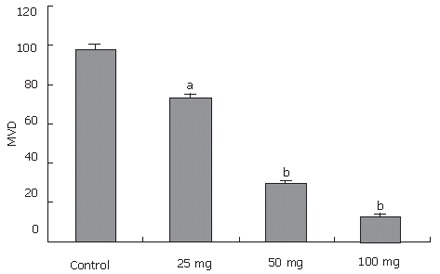
Figure 5.

Tumors resected from mice at the experimental end point. Resected tumors were sectioned and stained as described in the text. Microvessel densities (MVD) were determined by counting CD34-positive endothelial cells in the sections and presented as mean ± SE positive cells/field from the three “hot-spot” fields. aP < 0.05, bP < 0.01 vs control group.
