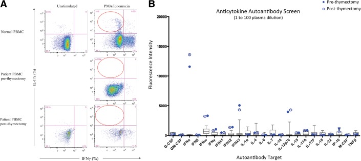
Figure 2.
Evidence of Th17 deficiency by flow cytometry and autoantibodies. (A) IL-17a production (%) in CD4+ memory T cells (red circles) for normal (top panels) and patient peripheral blood mononuclear cells pre- and postthymectomy (middle and lower panels, respectively). Unstimulated (left panels) or stimulated with phorbol 12-myristate 13-acetate/ionomycin (right panels) conditions. (Right side of each panel shows interferon [IFN] γ–producing CD4+ memory T cells.) Unstimulated condition not available for prethymectomy sample because of lymphopenia. B) Anticytokine autoantibodies before and after thymectomy. Plasma was mixed with the cognate beads, washed, and tested against human IgG.

