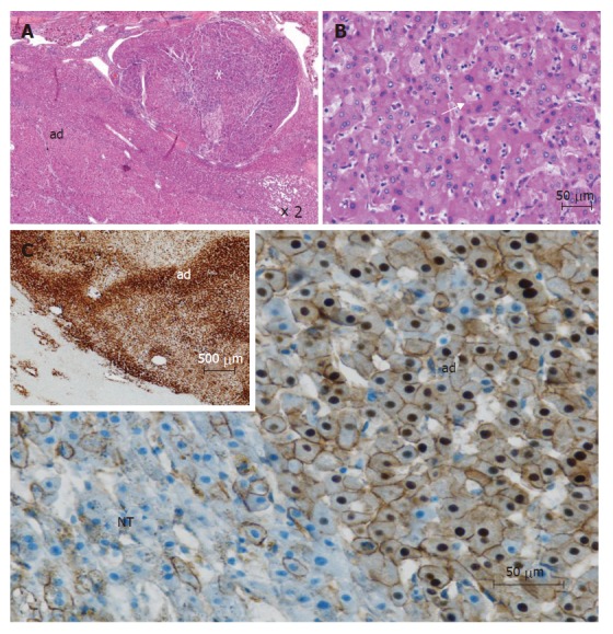
Figure 2.

β-catenin mutated adenomas (from different cases). A: This tumor is composed of 2 parts: an adenoma (ad) and a hepatocellular carcinoma (white asterix) (HE); B: In this adenoma, some irregular nuclei and acinar arrangements are seen (white arrow) (HE); C: immunohistochemistry: cytoplasmic and nuclear overexpression of β-catenin in adenoma (ad), contrasting with normal membranous staining in adjacent non tumoral liver (NT); Inset (upper left): homogeneous immunostaining with glutamine synthetase in adenoma (ad), contrasting with normal staining of pericentral hepatocytes (lower left).
