Figure 2.

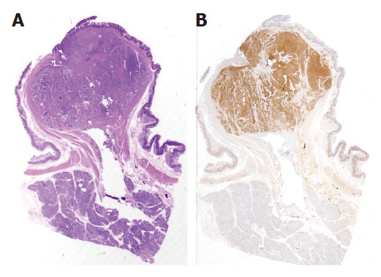
Low-grade microscopic view of the surgical specimen showing a well-differentiated endocrine carcinoma in the papilla major (A) and a strong immunohistochemical expression of synaptofisin (B).

Low-grade microscopic view of the surgical specimen showing a well-differentiated endocrine carcinoma in the papilla major (A) and a strong immunohistochemical expression of synaptofisin (B).