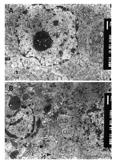
Figure 1.

Mitochondria appearance under electron microscope (EM × 6000); A: Mitochondria in normal group; B: Mitochondria in model group. M: mitochondria, G: glycogen, N nucleus, ER: endoplasmic reticulum, LD: lipid droplet. The long arrow shows abnormally distributed chromatin in nuclei, the short one is megamitochondrion and the arrow head is U-type mitochondria.
