
Figure 2.
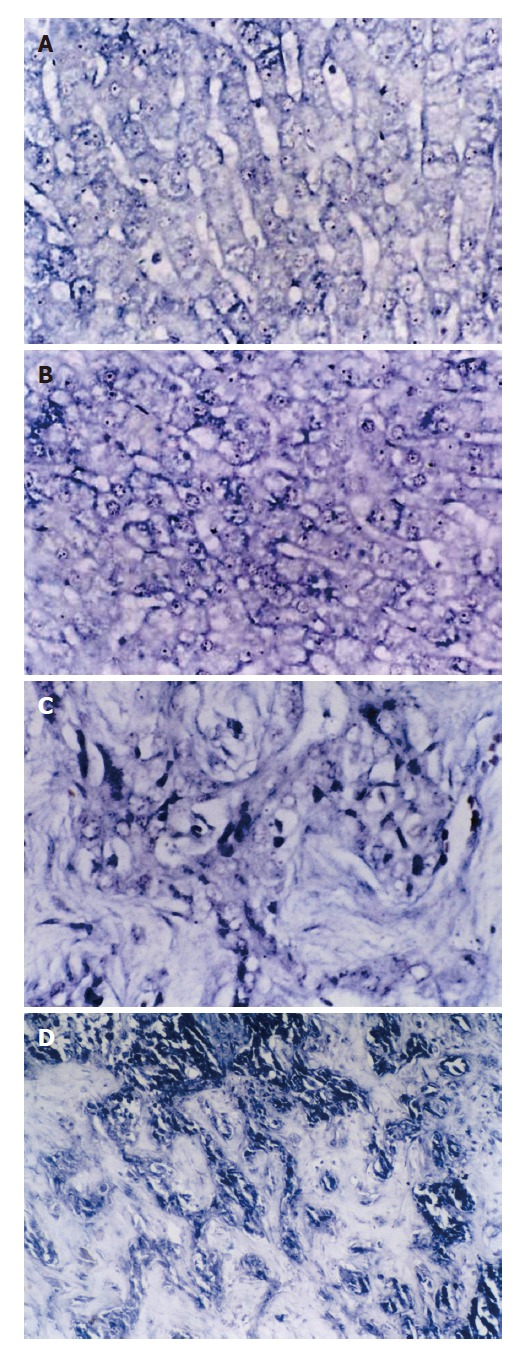
SePmRNA expressions in liver tissues. A: In normal liver tissues, the positive signals were in nucleus and cytoplasm, mostly in nucleolus, and the stained granules are large in nucleolus and around nucleus (ISH × 400); B: In liver cirrhosis tissues, the positive signals were mainly in nucleolus and cytoplasma, and the signals around nucleus and inner nucleus were less than in normal liver tissues (ISH × 400); C: In HCC tissues, the positive signals were in the cytoplasm, but less in nucleus. (ISH × 400); D: In hepatic interstitial substance, the positive signals were in the matrix, mainly in vascular endothelial cells and lymphocytes of vasculature (ISH × 400).