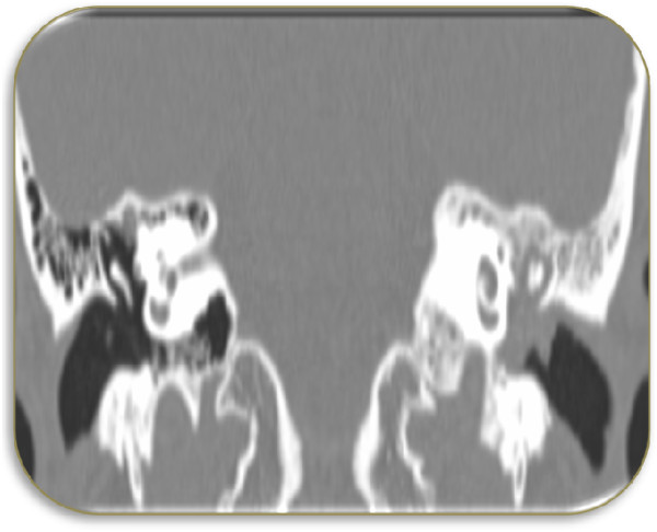
Figure 2.

Computed tomography scan of inner part of left external auditory canal. An irregular soft tissue lesion seen in the inner part of the left external auditory canal just lateral to the tympanic membrane, soft tissue density is also seen implicating the Prussak’s space, epitympanum, mesotympanum and hypotympanum.
