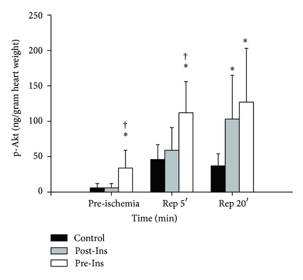
Figure 6.

Time-course changes of p-Akt before and after ischemia in the three groups. Muscle samples for p-Akt concentrations (ng/gram of dry heart weight) were taken after 20 min of preconditioning (preischemia, n = 12 in each group) and after 5 min of reperfusion (n = 12 in each group) and 20 min of reperfusion (n = 12 in each group). The data are presented as means ± SD. *P < 0.05 versus Control; † P < 0.05 versus Post-Ins. In Pre-Ins, p-Akt increased after administration of insulin before ischemia (preischemia). After 5 minutes of reperfusion, p-Akt was elevated in the Pre-Ins when compared with Control and Post-Ins (Rep 5′). The p-Akt levels in the Pre-Ins were still higher than Control after 20 minutes of reperfusion (Rep 20′).
