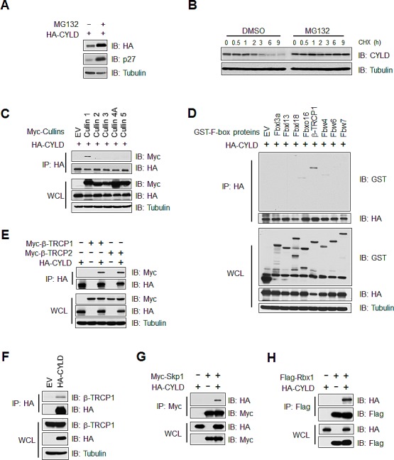
Figure 1. CYLD interacts with the SCFβ-TRCP E3 ligase complex.

(A) Immunoblot (IB) analysis of whole cell lysates (WCL) derived from DLD1 cells transfected with HA-CYLD with or without MG132 (15 μM) treatment. (B) RAW264.7 mouse macrophage cells were treated with RANKL (50 ng/ml) for 24 hours and then treated with or without MG132 (15 μM) for 6 hours prior to cycloheximide (CHX) treatment (20 μg/ml). At the indicated time points, WCL were prepared, and IB analysis was performed with the indicated antibodies. (C) IB analysis of WCL and anti-HA immunoprecipitates (IP) derived from 293T cells, in which HA-CYLD was co-transfected with empty vector, Myc-Cullin1, 2, 3, 4A or 5 expression plasmids. (D) IB analysis of WCLs and HA-IP derived from 293 cells transfected with HA-CYLD and the indicated GST-tagged F-box proteins. (E) IB analysis of WCL and anti-HA IP derived from 293T cells, in which HA-CYLD was co-transfected with Empty vector (EV), Myc-β-TRCP1 or Myc-TRCP2 plasmids as indicated. (F) IB of WCLs and anti-HA IP derived from 293T cells transfected with HA-CYLD or EV as a negative control. (G) IB analysis of WCL and anti-Myc IP derived from 293T cells transfected with HA-CYLD and Flag-Skp1 as indicated.(H) IB analysis of WCL and anti-Flag IP derived from 293T cells transfected with HA-CYLD and Flag-Rbx1 as indicated.
