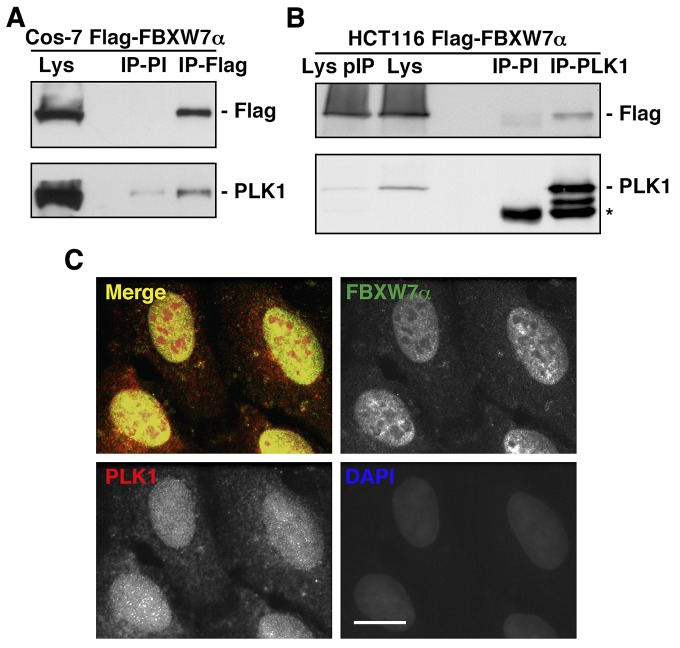
Figure 1. FBXW7α and PLK1 interact in the nuclei of mammalian cells.
(A) Cos-7 cells were transiently transfected with pCDNA3.1-Flag-FBXW7α and nuclear extracts immunoprecipitated with anti-Flag monoclonal antibody or normal mouse serum (PI). Immunoprecipitates materials were analyzed by Western blotting. Lys: nuclear extracts from Cos-7 transfected cells. (B) Whole cell extracts from HCT116 transfected cells were used to immunoprecipitate PLK1, and complexes were analyzed by immunoblotting. IP-PI: immunoprecipitation with normal mouse serum. Lys and Lys pIP: whole cell extracts from HCT116 transfected cells before (Lys) and after (Lys pIP) PLK1 immunoprecipitation. Asterisk indicates IgG heavy chains. (C) U2OS cells were stained for FBXW7α, PLK1 and DNA. In the merge, FBXW7α staining is shown in green, PLK1 in red, and DAPI in blue. Bar, 10μm.

