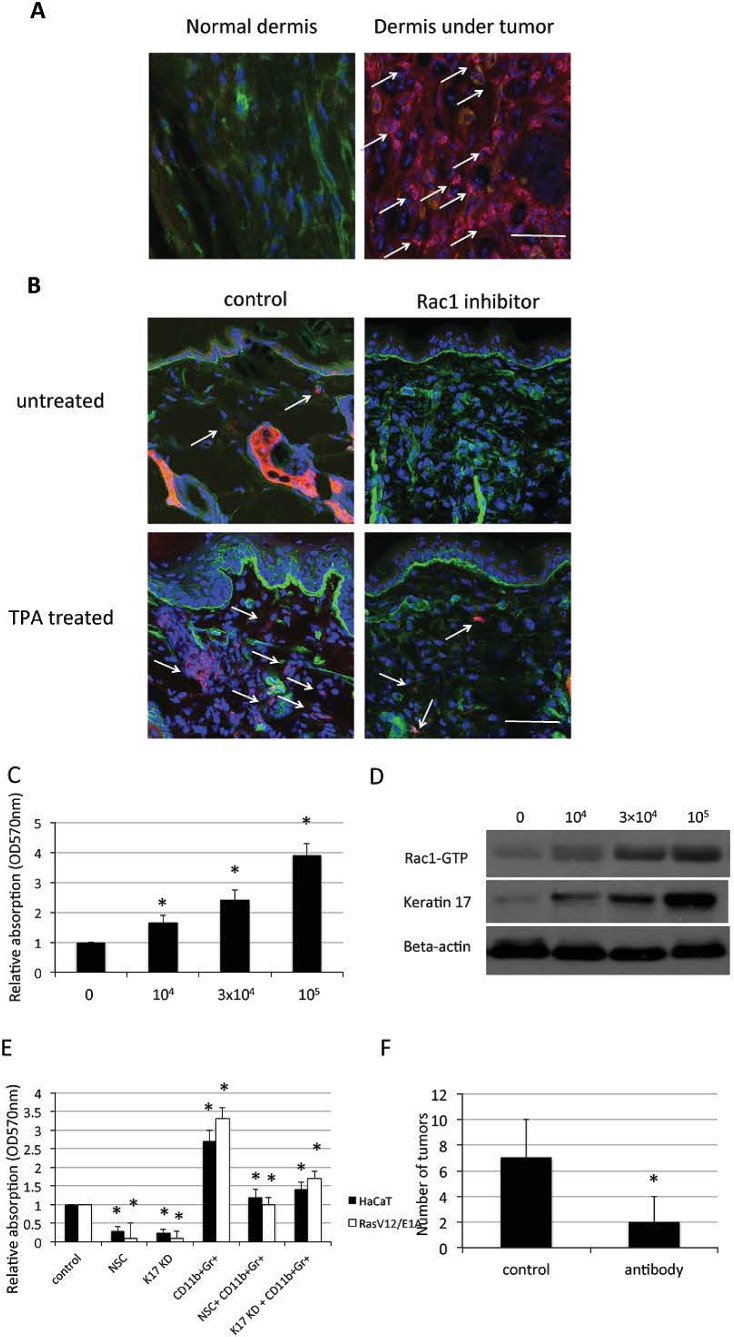
Figure 5. CD11b+Gr1+ cell infiltration and keratinocyte proliferation through Rac1 and keratin 17.
A, Immunofluorescence for MPO positive MDSC cells (red) in the dermis of SCC patients. α6 (green), DAPI (blue) counterstaining indicates nuclei. Scale bar: 20 μm. B, 8 week old mice were treated with Rac inhibitor NSC23766 at 10mM 30min prior to TPA treatment or treated with TPA only, 48h later mice were sacrificed. Immunofluorescent staining was performed for Gr1+ cells (red) and α6 (green) in skin. DAPI (blue) counterstaining indicates nuclei. Scale bar: 20 μm. C, HaCaT cells were cocultured with the indicated number of CD11b+Gr1+ cells in 96-well plates for 72 h and proliferation of HaCaT cells was measured by crystal violet assay. Assays were performed in triplicate. The means with SD are shown (n=8 replicates/group). * p<0.01 compare to control. D, HaCaT cells were cocultured with the indicated number of CD11b+Gr1+ cells in 6-well plates for 24 h and proteins were analyzed by western blot for keratin 17. Rac1-GTP was evaluated using pull-down assay. E, HaCaT cells or RasV12/E1A-transfected keratinocytes were treated with Rac1 inhibitor NSC23766 (NSC), small hairpin RNA against keratin17 (KD), or cocultured with 104 CD11b+Gr1+ cells in 96-well plates for 72 h with proliferation measured by crystal violet assay. F, Control (con) mice and mice treated by intraperitoneal injection with Gr1 monoclonal antibody every four days for 4 weeks. TPA was used treated mice for 2 weeks (n = 10). Shown is average number of tumors per mouse. * p<0.01 compare to control.

