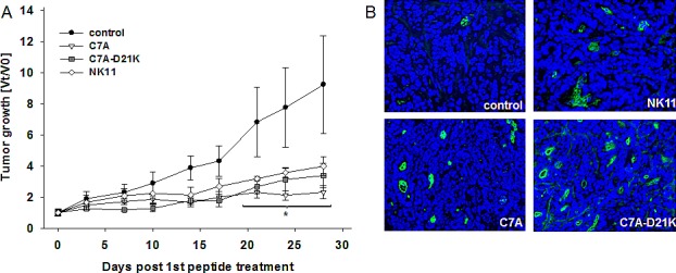
Figure 7. In vivo growth kinetics and tumor cell apoptosis of HROC24 tumors in NMRI Foxn1mice with or without local treatment.

(A) Therapy was performed by repetitive local application of HDPs C7A or C7A-D21K (1 mg/kg bw) every third day (a total of 9 injections) (n=5 per group). Tumor-carrying control animals received either equivalent volumes of peptide NK11 or saline (n=5 per group). Tumor volumes are given as x-fold increase vs. day 0 (Vt/V0) (start of treatment) ± SD. *p<0.05 vs. PBS. (B) Representative immunofluorescence staining of apoptotic cells within HROC24 tumors. Cryopreserved tumor sections were stained with anti-M30 CytoDeath antibody, followed by anti-mouse IgG FITC antibody and nuclear DAPI as described in material and methods.
