
Figure 2. DU-145 cells express receptors of LHRH on the cell membrane.
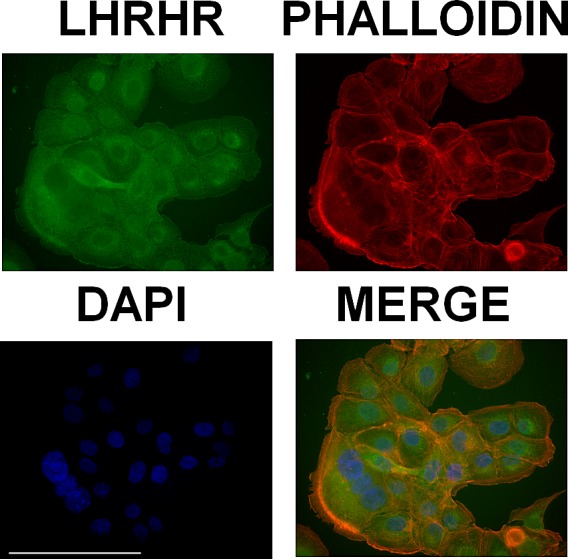
LHRH receptors were labelled by using immunocytochemistry (green), the actin cytoskeleton was visualized by TRITC-conjugated phalloidin (red) and nuclei were stained with DAPI (blue). Scale bar corresponds to 100 μm.