
Figure 10.
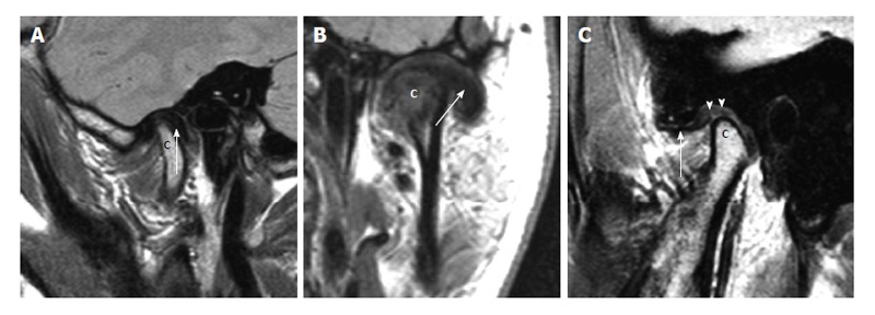
Other types of disk displacement. A: Posterior disk displacement. Sagittal proton density weighted magnetic resonance imaging (MRI) in the closed mouth position demonstrates posterior displacement of the disk (arrow) in relation to the mandibular condyle (the letter, c); B: Lateral disk displacement. Coronal proton density weighted demonstrates lateral displacement of the disk (arrow) in relation to the mandibular condyle (the letter, c); C: Pseudodisk. Sagittal proton density weighted MRI in the closed mouth position demonstrates anterior displacement of the disk (arrow) in front of the mandibular condyle (the letter, c). The thickening of the posterior attachments (arrowheads) superior to the mandibular condyle is seen as “pseudodisk”.