
Figure 2.
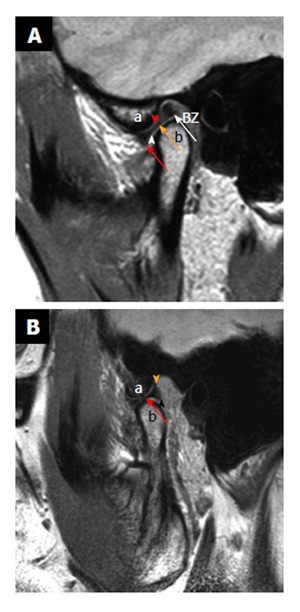
Normal anatomy. Sagittal proton density weighted closed mouth and open mouth view of magnetic resonance imaging. A: On the closed mouth view, the disk is located posterior to the articular eminence (the letter, a). It can be noted that the “bow-tie” shape of the disk: Thicker anterior band (red arrow) and posterior band (white arrow) with a thinner central zone (orange arrow). Bilaminar zone (BZ) is located posterior to the posterior band. It can also be noted that the inferior joint compartment (white arrowhead) between the disk and the mandibular condyle (the letter, b) and superior joint compartment (red arrowhead) between the articular eminence and the disk; B: On the open mouth view (in a different patient), the thinner intermediate zone (red arrow) of the disk is interposed between the articular eminence (the letter, a) and the condylar head (the letter, b) in a “bow-tie” fashion. Orange arrowhead demonstrates temporal lamina and black arrowhead indicate inferior lamina.