
Figure 3.
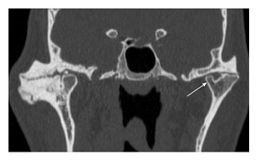
Bifid condyle. Coronal reformatted computed tomography image through the temporomandibular joint (TMJ) demonstrates bifid left mandibular condyle. It can be noted that one of the condyles (arrow) is smaller than the other. Advanced degenerative changes are noted in bilateral TMJ.