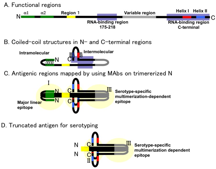
Figure 4.
A schema of trimerized hantavirus N. A. Functional regions of N were plotted on the primary structure of hantavirus N shown in Figure 1, Figure 2, and Figure 3. B. Interactions in the N-terminal and C-terminal part of hantavirus N. C. Antigenic regions mapped by poly- and monoclonal antibodies: From the competitive binding assay, two major antigenic regions were found in HTNV N. One was the N terminus (antigenic region I) and the other was the C terminus (antigenic region III). The central region was not a major antigenic site. Serotype-specific epitopes were found in the edge region and as discontinuous epitopes [26]. D. Concept of serotyping antigen based on truncation of N antigen. By deletion of group-common and major linear epitopes, serotyping antigens were designed [55].

