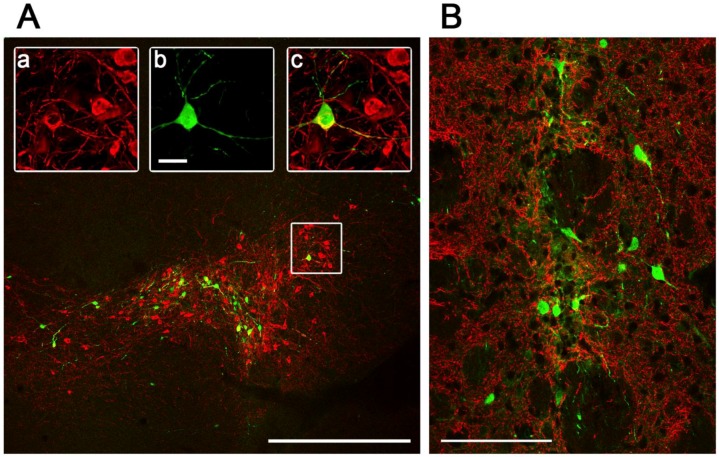
Figure 5.
Pattern of Ad5-CGW-CK2-mediated GFP expression at the site of stereotactic delivery. Confocal immunohistochemical analysis of GFP expression by Ad5-CGW-CK2 stereotactically delivered to mouse SN (A) and STR (B) results in striking neuronal tropism. (A) Low-power image of the injected SN. Only neurons are positive for GFP expression. (a,b,c) Magnification of the region outlined in (A). A TH (red, a)/GFP (green, b) double-positive neuron is clearly visible surrounded by uninfected dopaminergic (DA) neurons (merge, c). (B) When Ad5-CGW-CK2 is delivered to the STR, GFP-expressing cells with neuronal morphology are observed. Despite thick (40 µm) sections and assessment of numerous slices around the injection site, no non-neuronal GFP expressing cells could be appreciated in the brains of animals receiving Ad5-CGW-CK2. Red = TH. Green = GFP. Bar in A = 500 μm. Bar in b (for a, b, c) = 2.5 μm. Bar in B = 100 μm.

