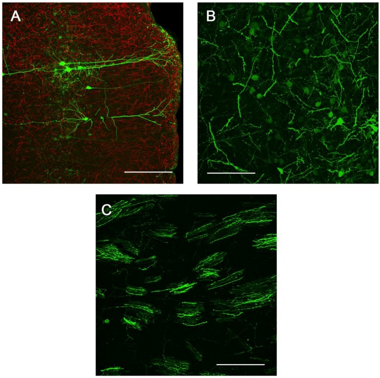
Figure 6.
Sites of presynaptic GFP expression resulting from stereotactic delivery of Ad5-CGW-CK2 to SN. Regions with afferent projections to the SNc showed large numbers of neurons infected by Ad5-CGW-CK2. Regions presynaptic to the site of injection, including pyramidal neurons of the lateral cortex (A) and neurons of the globus pallidus (B), showed robust GFP expression. Additionally, GFP-positive fibers could be traced coursing through the STR in bundled myelinated fibers (C, correlating to the human internal capsule). All images are flattened confocal z-stacks through the regions indicated. Green = GFP. Red = TH. Bar in A = 200 µm, Bar in B and C = 100 µm.

