
Fig. 2.
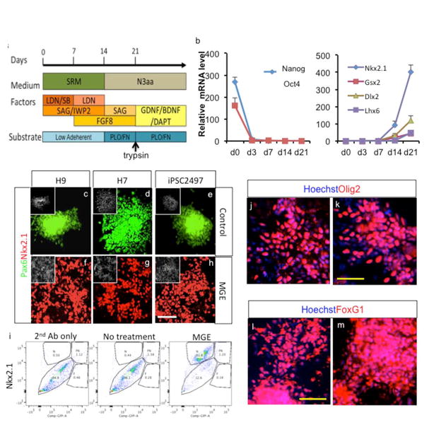
Optimized MGE derivation protocol efficiently generates MGE cells from multiple PSC lines. a. Overview of optimized MGE derivation protocols. b. Gene expression analysis during MGE derivation of H9 cells, assayed by real time PCR (Mean ± S.E.M.; n=3). c-h. Combined and temporal treatment with IWP2, SAG and FGF8 results in robust induction of MGE cells from H9 and H7 hESCs as well as iPSC2497, assayed after 25 days of differentiation. White scale bar: 100μm. i. FACS analysis of MGE generation of H9 cells after Nkx2.1 staining. j-m. Derived cells highly express independent ventral telencephalic marker Olig2 and telencephalic marker FoxG1, assayed after 25 days of differentiation. Yellow scale bar: 50μm.