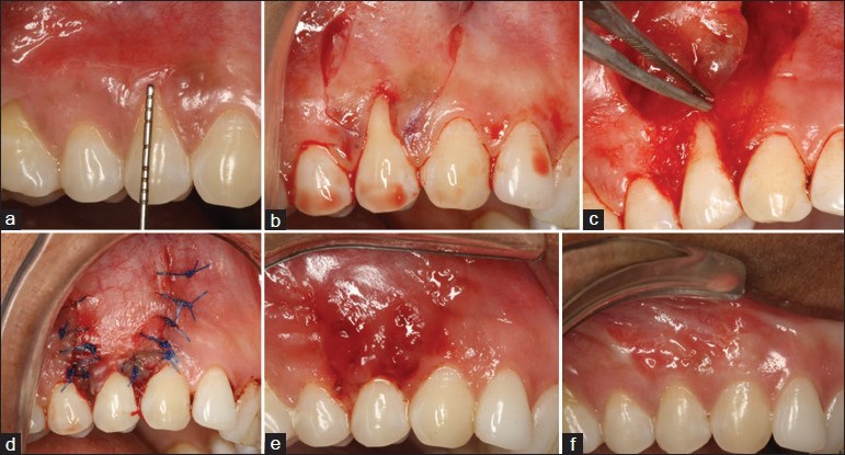
Figure 1.

(a) Preoperative gingival recession (3 mm), (b) oblique and releasing incisions given, (c) recipient bed prepared, amnion allograft being placed, (d) flap coronally advanced and sutured, (e) healing after 10 days and (f) 6 months postoperative healing showing complete root coverage and esthetic color match
