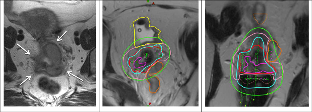
Fig 1.
Patient with FIGO stage IIIB treated with EBRT and 2 fractions of PDR MRI-guided brachytherapy. Left panel shows transverse MRI at time of diagnosis with parametrial proximal involvement (left) and to the pelvic wall (right). At time of brachytherapy there was still residual parametrial disease (left proximal and right distal). A combined intracavitary/interstitial applicator (5 needles) was used. Middle and right panel show para-transverse and coronol MRI at time of brachytherapy with the applicator in situ. The volumes are: residual GTV (magenta), CTVHR (red), CTVIR (pink), bladder (yellow) and sigmoid (orange). Isodoses 15 Gy (cyan) and 7.5 Gy (green) correspond to 84 Gy and 60 Gy in terms of total EBRT and brachytherapy EQD2.

