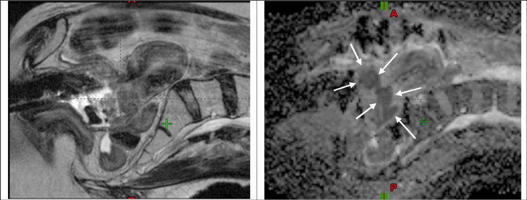
Fig 2.
MRI at time of brachytherapy for a locally advanced cervical cancer patient with stage IB2 disease treated at Dept of Radiation Oncology, Washington University, St. Louis. Left and right panel show sagittal T2w and ADC images, respectively, obtained at 4. fraction of brachytherapy with the intracavitary applicator in situ. At the time of imaging 13 fractions of IMRT had been delivered with an integrated midline blocking as well as 3 fractions of brachytherapy of 6.5Gy to point A. A significant residual GTV mass is clearly identified in the cervix region on the ADC map (arrows). The bright signal regions on the T2w image indicate residual GTV, but appear with less clear borders towards the surrounding tissue as compared with the ADC map.

