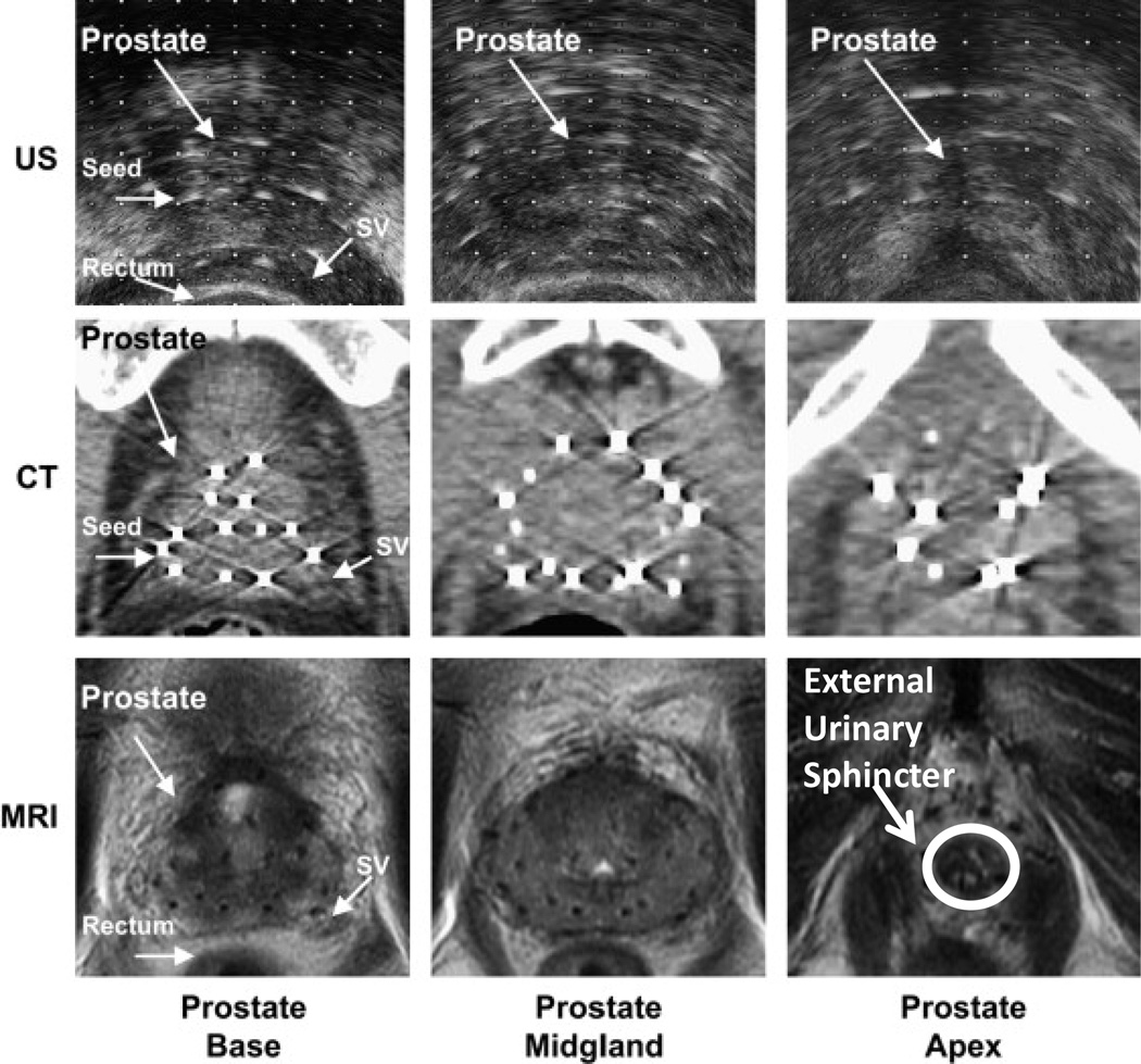
Fig 3.
Ultrasound (US), CT, and MRI images of the base, midgland, and apex of the prostate following LDR brachytherapy. MRI has superior soft tissue delineation of the prostate over ultrasound and CT. Urinary irritation and bother symptoms, which are more common in prostate brachytherapy, may be reduced with better anatomic delineation of the external urinary sphincter (indicated in bottom right) during simulation and treatment planning. Additionally, better anatomic delineation of the apex, base, neurovascular bundles, bladder neck, and intraprostatic ejaculatory ducts may also improve disease outcomes and reduce treatment related morbidity.

