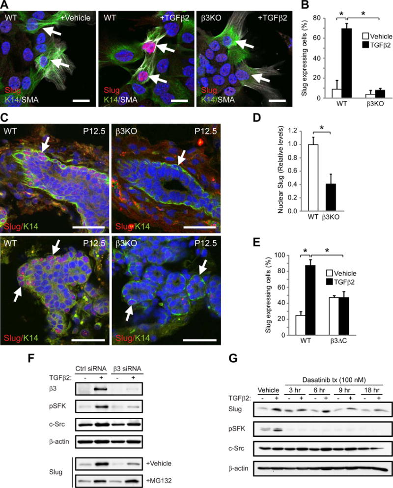
Figure 6. β3 is required for Slug activation in response to TGFβ2 or pregnancy.

(A,B) Slug expression in K14+SMA+ cells from virgin WT and β3KO mammary cells stimulated with vehicle or TGFβ2. (A) Representative images of Slug expression in K14+SMA+ cells (arrows). Scale bars, 20 μm. (B) Quantitation of the percentage of Slug+K14+SMA+ cells. P=0.0439 (vehicle versus TGFβ2 in WT cells) and P=0.0342 (WT versus β3KO cells stimulated with TGFβ2). (A,B) WT, n=3, β3KO, n=3.
(C,D) Slug expression in WT and β3KO P12.5 mammary glands. (C) Representative images of Slug in K14+ cells (arrows). Scale bars, 20 μm. (A,C) Nuclei are stained blue in all panels. (D) Histogram showing the relative levels of nuclear Slug expression. Data for each mouse represents the average nuclear Slug expression from 5 fields normalized to total nuclear stain. P=0.0113. (C,D) WT, n=8, β3KO, n=6.
(E) Quantitation of the percentage of Slug-expressing K14+SMA+ cells from virgin WT and β3ΔC mammary cells stimulated with vehicle or TGFβ2. WT, n=2, β3ΔC, n=2. P=0.029 (vehicle versus TGFβ2 in WT cells) and P=0.032 (WT versus β3ΔC cells stimulated with TGFβ2). (B,D,E) Data represent the mean ± s.e.m. and statistical analysis performed by Student’s T-tests. *P<0.05.
(F,G) Immunoblots of MCF10A cells stimulated with TGFβ2 or vehicle control and probed for the indicated proteins. (F) Cells were transfected with Control (Ctrl) or β3 siRNA and additionally treated with vehicle or proteasome inhibitor (MG132) for 5 hr prior to lysis. (G) Cells were treated with 100 nM Src inhibitor (Dasatinib) for the indicated length of time prior to lysis. (F,G) Data shown is representative of 3 independent experiments. See also Figures S6 and S7.
