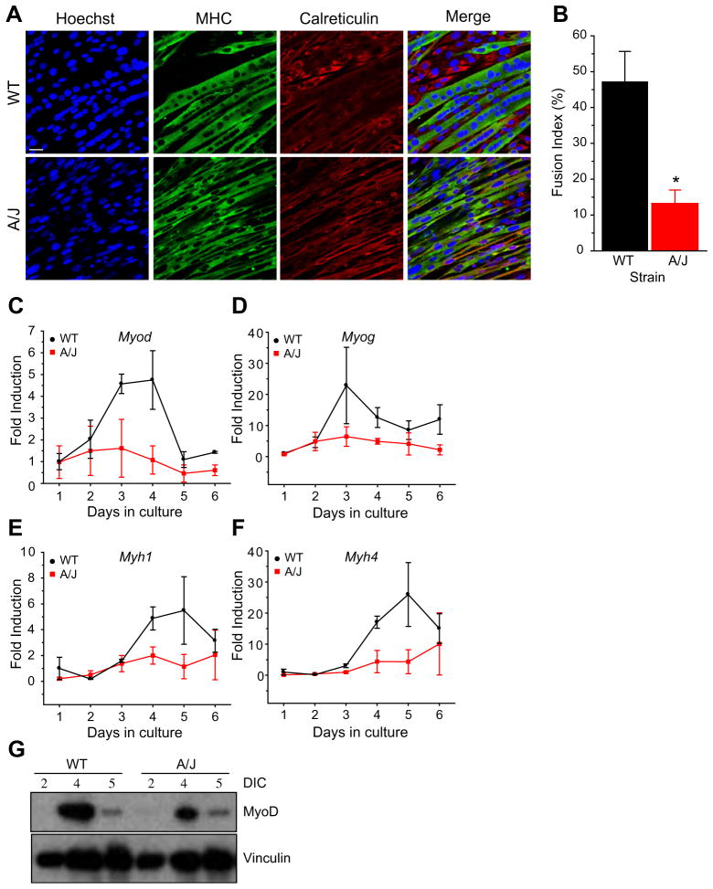
Fig. 3.
A/J myotubes form thinner myotubes and show a reduced myofusion index. (A) Immunostaining of A/J myotubes with antibodies for myosin heavy chain (MHC), calreticulin and Hoechst. (B) Quantitation of the fusion index. n = 386 (WT), 448 (A/J) total nuclei were counted containing 186 (WT) and 63 (A/J) myotubes in four non-overlapping fields. P < 0.05. (C–F) Real-time expression of myogenic genes during the time course of differentiation. Cultures from three independent H-2K A/J or WT clones were differentiated for up to 6 days in culture. Myogenic gene expression was measured using qPCR for MyoD (Myod) (C), myogenin (Myog) (D), myosin heavy chain 1 (Myh1) (E) and myosin heavy chain 4 (Myh4) (F). (G) Top: Western blot of MyoD expression between 2 and 5 days in culture (DIC) in WT and A/J H-2K cells. Bottom: Vinculin was used as a protein loading control. Scale bar = 25 μm.

