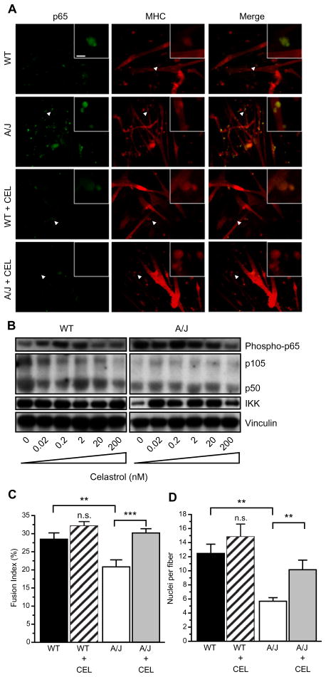
Fig. 4.
Celastrol blocks NFκB signaling. (A) Celastrol inhibits p65 activation and increases fiber diameters. Myoblasts were cultured in 6-well dishes. Twenty-four hours later, media was switched to differentiation media containing 200 nM celastrol. After 5 days in culture, myotubes were stained with antibodies to phospho-p65 (green) and anti-MHC (red). Phospho-p65 levels were higher in cultures of A/J cells compared to wild-type cells. Incubation of differentiating myoblasts with celastrol decreased phospho-p65 in the cultures and increased diameters of cells, shown with MHC staining. Inset shows higher magnification of area indicated by arrowhead. Scale bar = 20 and 5 μm in inset. (B) Cells were plated in 6-well dishes and celastrol was added at indicated concentrations after 24 h, the myotubes were harvested after 5 days. Western blot showing that phosphorylated p65 subunit of NFκB is upregulated in A/J cells and decreases with celastrol treatment (top). Total expression of p105, p50, and IKK is not altered with celastrol treatment. (C and D) Fusion index and nuclei per fiber were calculated from six random fields in two independent experiments. Celastrol significantly increased fusion index (C) and nuclei per fiber (D) in A/J cultures. Scale bar = 20 μm. **P < 0.05. ***P < 0.01.

