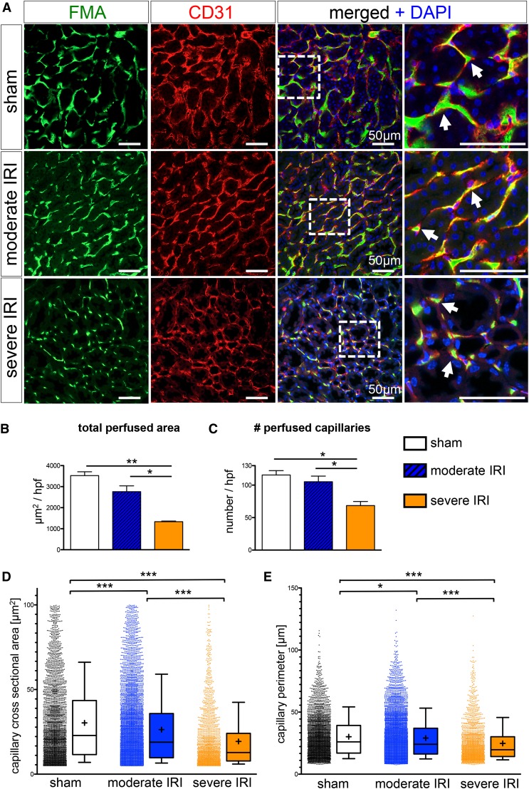
Figure 3.
High-throughput software–based analysis of fluorescence microangiography reveals reduced capillary number and caliber after severe IRI. (A) FMA after sham surgery and moderate and severe IRI, together with CD31 immunostaining, demonstrates capillary rarefaction in response to severity of injury (arrows indicate capillaries with red CD31+endothelial cells surrounding the green FMA solution; all scale bars are 50 µm). (B and C) Severe IRI results in a significant reduction of the total cortical cross-sectional capillary area per high-power field [×400, inner cortex] (B) and a significant reduction in capillary number (C). (D and E) The cortical individual capillary cross-sectional area (D) (mean±SEM, sham: 30.31±0.42 µm2; moderate IRI: 26.29±0.28 µm2; severe IRI: 19.34±0.38 µm2) and perimeter (E) (sham: 30.19±0.31 µm; moderate IRI: 29.13±0.22 µm; severe IRI: 24.77±0.35 µm) was significantly reduced after both moderate and severe IRI. (Of note, data represent n=3 mice in the sham group and severe IRI group and n=6 mice in the moderate IRI group; mean±SEM in B and C; box and whiskers with 10th–90th percentiles in D and E; + indicates mean in D and E. *P<0.05; **P<0.01; ***P<0.001, one-way ANOVA with post hoc Bonferroni correction). DAPI, 4′,6-diamidino-2-phenylindole.

