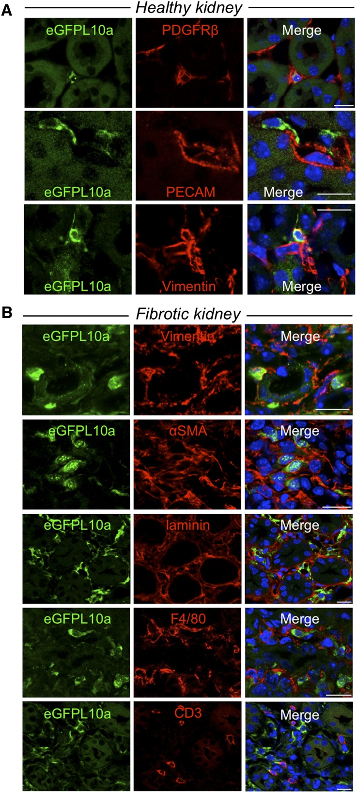
Figure 3.
Immunostaining demonstrates cell type–specific expression of eGFP-L10a in Peri/FibroTRAP kidney. (A) In uninjured kidney, eGFP-L10a+ cells (green) of kidney medulla stain positive for pericyte/fibroblast markers PDGFR-β and vimentin and are closely associated with endothelial cell marker platelet endothelial cell adhesion molecule (PECAM)+peritubular capillaries, consistent with a pericyte/fibroblast identity. (B) In fibrotic kidney (5 days UUO), eGFP-L10a+ cells remain strictly confined to the tubulointerstitial compartment, as documented by antilaminin staining, continue to express vimentin but also express the myofibroblast marker αSMA, suggesting a pericyte/fibroblast to myofibroblast transformation. Although spatially close, eGFP-L10a+ cells do not costain with macrophage marker F4/80 or T-cell marker CD3. Scale bars: 10 µm.

