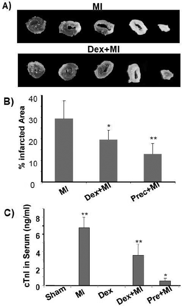
Fig. 1. Dexamethasone reduces myocardial infarct size and cTnI release.
C57BL6 mice at 8 weeks old were used for left anterior descending coronary artery occlusion surgery. For preconditioning, the coronary artery was occluded for 5 mins then released for 5 mins for two cycles. Dexamethasone (i.p. 20 mg/kg) was administered 20 h prior to the surgery. At 24 h after permanent occlusion, the hearts and plasma were collected for triphenyl tetrazoliumchloride staining to measure areas of infarction (A, B) and for serum cTnI concentration assay respectively. A serial of transverse sections of representative left ventricles downstream of ligature were shown (A). Total left ventricular areas or areas of infarct were quantified by tracing using NIH Image J software. cTnI was not detectable in sham operated or dexamethasone treated control groups. The data represent means + standard deviations from 3 animals in each group (B). * indicates p<0.05 and ** indicates p<0.01 when compared to MI (B) or sham operated control (C).

