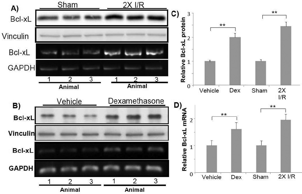
Fig. 4. Dexamethasone causes elevated expression of Bcl-xL in the myocardium.
C57BL6 mice at 8 weeks old were used for left anterior descending coronary artery occlusion surgery or dexamethasone treatment. For preconditioning, the coronary artery was occluded for 5 mins then released for 5 mins for two cycles. Ventricular tissues were collected immediately at the end of the ischema-reperfusion cycles for Western blot analyses (80 µg protein/lane) or RT-PCR (A). Dexamethasone (i.p. 20 mg/kg) was administered 20 h prior to ventricular tissue collection (B). Each lane represents Bcl-xL protein or mRNA level from one animal. The quantitative Bcl-xL protein data (C) or mRNA (D) show means + standard deviations of band densities from three animals. Vinculin (A) or GAPDH (B) was used as a loading control. * indicates p<0.05 and ** indicates p<0.01.

