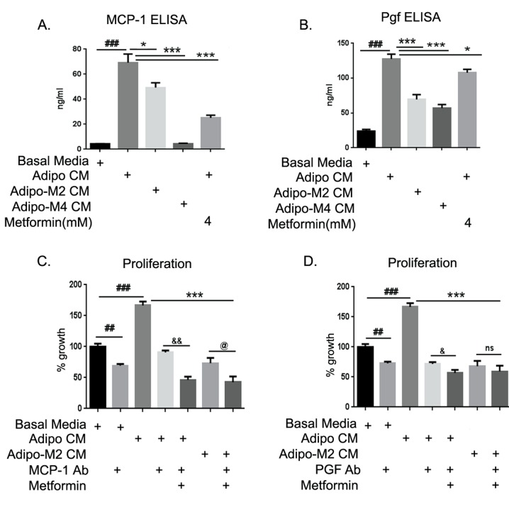
FIGURE 7. MCP-1 and Pgf neutralization blocked adipocyte potentiation of ID8 proliferation.
ELISA (enzyme-linked immunosorbent assay) for MCP-1 (monocyte chemo-attractant protein-1) and Pgf (placental growth factor) were performed in conditioned media (CM) from mature adipocytes and in supernatants from ID8 cells treated with various CM in absence or presence of metformin. Subtraction values from both ELISA readings were taken as quantitative concentration of cytokines being released by ID8 cells. (A) MCP-1 (###p < 0.001 compared to basal media; *p < 0.05, **p < 0.01, ***p < 0.001 compared to adipocyte CM [adipo CM]). (B) Pgf (###p < 0.001 compared to basal media; *p < 0.05, ***p < 0.001 compared to adipo CM). (C) ID8 cells plated in 96-well plate were exposed to the various CM in presence of the indicated treatments of MCP-1 antibody or Pgf antibody (D) with or without metformin, and increase in proliferation was estimated by MTT assay performed at 48 hours (##p < 0.01, ###p < 0.001 compared to basal media; ***p < 0.001 compared to adipo CM; &p < 0.05, &&p < 0.01 metformin and antibody combination compared to antibody alone in presence of adipo CM; @p < 0.05, ns: non-significant metformin and antibody combination compared to CM from adipocytes differentiated in presence of metformin 2mM [adipo M2 CM]).

