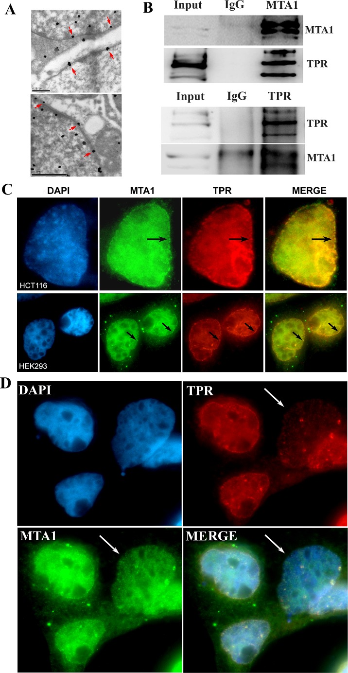
Figure 5. MTA1 interacts and colocalizes with TPR at the nuclear envelope.
A. The localization of MTA1 on the nuclear envelope (indicated with red arrows) using immuno-electron microscopy; B. Co-IP analysis of the interaction between MTA1 and TPR; C. Immuno-colocalization of MTA1 and TPR at the nuclear envelope of HCT116 and HEK293 cells is indicated with black arrows; D. The knock down of TPR in HEK293 cells impairs the nuclear envelope localization of MTA1. The white arrow indicates a typical TPR knocked down cell.

