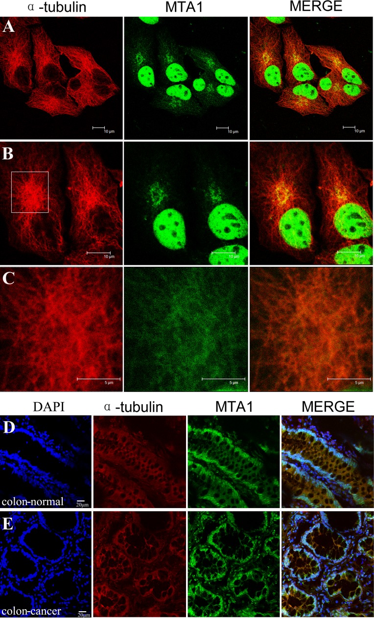
Figure 6. Localization of MTA1 on microtubules in the cytoplasm.
NCI-H446 cells and colon tissues were immuno-stained with specific antibodies against MTA1 and α-tubulin, followed by detection using a laser scanning confocal microscope. The localization of MTA1 on microtubules in NCI-H446 cells is shown in A, B, and C. An enlargement of the rectangular region in B is shown in C. D and E show the localization of MTA1 on microtubules in the cytoplasm of normal and cancerous colon tissues respectively.

