
Figure 2. Cells in the proximal regions of lin-41 gonads lose germline characteristics and differentiate into somatic cells, forming a teratoma.
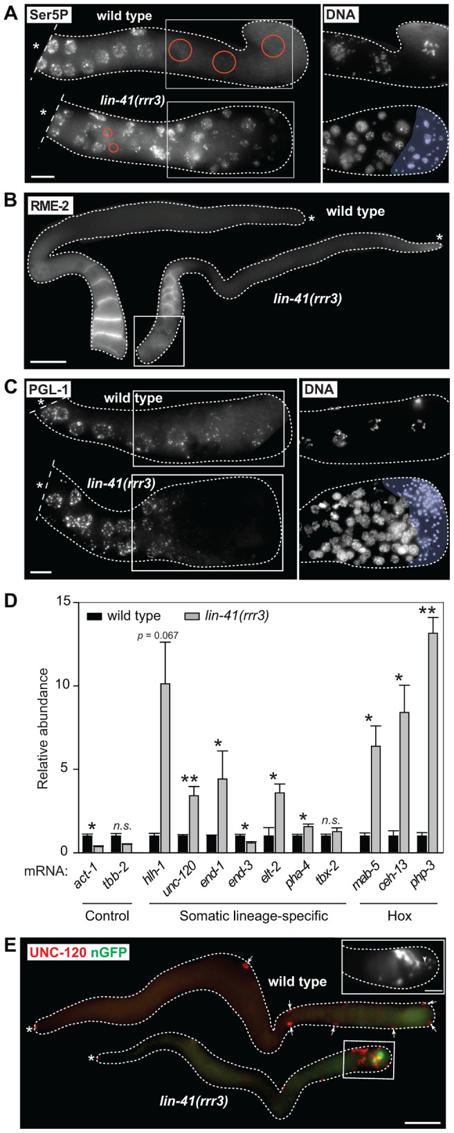
A. Fluorescent maximum intensity projections of gonads immunostained for the transcription-activating phosphorylation of Ser5 within the Pol II CTD (Ser5P). This phosphorylation was absent in the most-proximal wild-type gonads, but was present in all corresponding lin-41(rrr3) gonads (n>40). Encircled in red are nuclei containing low or no Ser5P. The corresponding DAPI-stained nuclei, from the boxed areas, are on the right. The proximal-most lin-41 gonad contains sperm (lightly colored in A and C), which explains the lack of Ser5P in this region. Scale bar: 10 µm. B. Fluorescent maximum intensity projections of gonads immunostained for an oocyte-expressed protein, the yolk receptor RME-2. In wild-type gonads, RME-2 is expressed in developing oocytes. In lin-41(rrr3) gonads, RME-2 is expressed in oocyte-like cells but is absent from the most proximal cells (boxed). Scale bar: 25 µm. C. Fluorescent maximum intensity projections of gonads immunostained for a germline-specific protein, PGL-1. PGL-1 (concentrated in RNA/protein granules) is present throughout the wild-type gonad but is eliminated in the proximal lin-41(rrr3) gonad (all gonads, n = 25). The corresponding DAPI-stained nuclei from the boxed areas are on the right. Scale bar: 10 µm. D. Detection of somatic cell-specific transcripts by RT-qPCR. “Somatic lineage-specific” indicates mRNAs expressed in somatic cell lineages: muscle (hlh-1, unc-120) or pharynx/gut (end-1, end-3, elt-2, pha-4 and tbx-2). “Hox” genes direct various aspects of somatic development. Each bar represents the mean of three independent biological replicates, the error bars represent the SEM and the significance of the differences has been calculated with the Student's t-test (“*”, p<0.05; “**”, p<0.01; “n.s.”, not significant). E. Fluorescent maximum intensity projections of gonads immunostained for the muscle-lineage marker UNC-120 and for a neuronal GFP reporter (nGFP). Arrows point to UNC-120-containing cells of the somatic gonad. In contrast to wild-type, UNC-120 and nGFP-expressing cells (boxed) are present within the lin-41(rrr3) germline. The inset shows nGFP-expressing cells from a different lin-41(rrr3) gonad, which extend long, neuronal-like processes (arrowhead). Scale bar: 50 µm.