
Figure 3. LIN-41 inhibits the transition from meiosis to mitosis in developing oocytes.
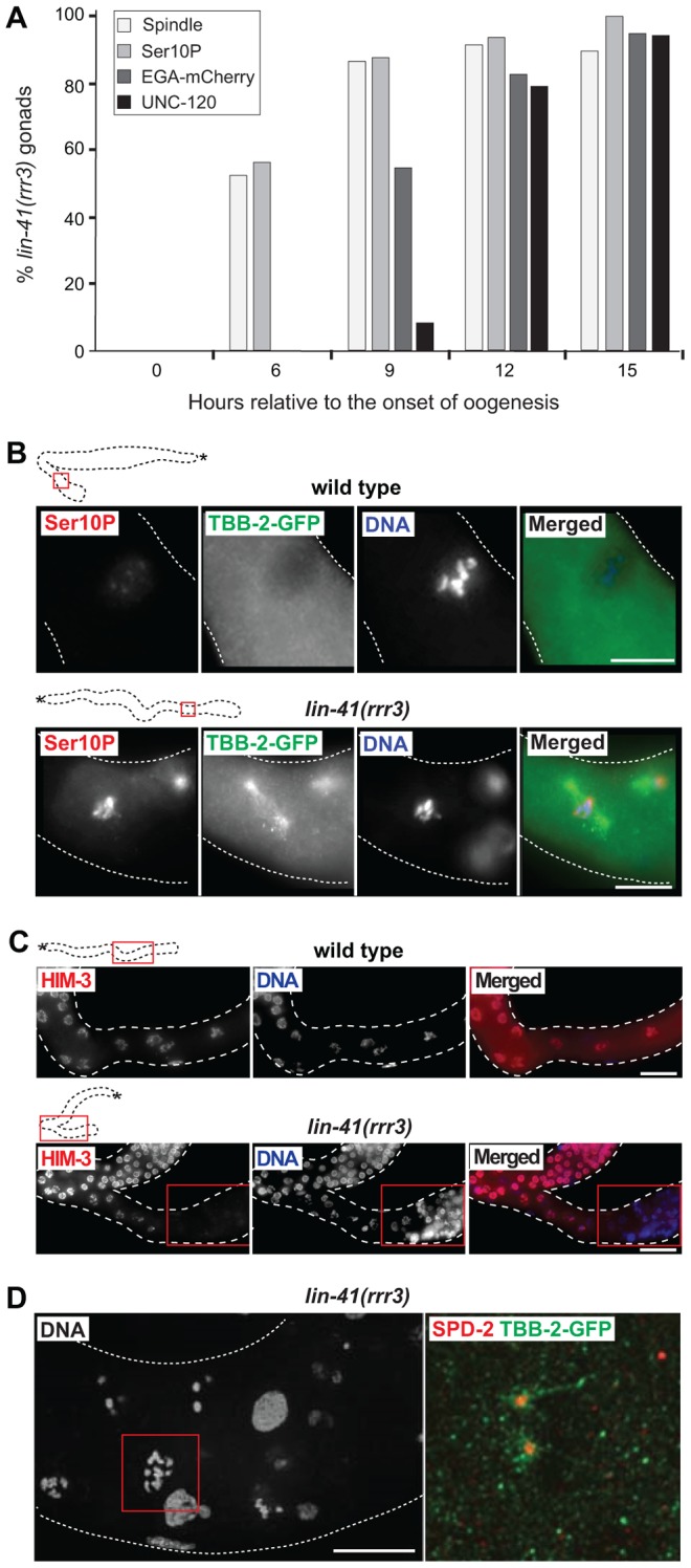
A. Time-course of EGA, mitotic chromosome condensation, spindle formation and somatic-like differentiation in lin-41(rrr3) gonads. The numbers indicate the fractions of lin-41(rrr3) gonads expressing EGA-mCherry (here the vet-4 promoter drives mCherry-tagged H2B) or assembling mitotic spindles (visualized with GFP-tagged β-tubulin; TBB-2-GFP), which was observed in live animals at the indicated time points. Additionally, these animals were immunostained for a mitotic marker (Ser10P) or UNC-120. At least 30 gonads were examined per each time-point/marker. B. Fluorescent maximum intensity projections of selected regions (boxed in red on the schematic gonads) of wild-type and lin-41(rrr3) gonads, immunostained for Ser10P and microtubule spindle (TBB-2-GFP), also stained by DAPI. Scale bars: 10 µm. C. Fluorescent maximum intensity projections of selected regions of wild-type and lin-41(rrr3) central-proximal gonads, immunostained for the meiotic marker HIM-3 and also stained by DAPI. Scale bars: 25 µm. D. Confocal images of maximum intensity projections of selected cells in the proximal lin-41(rrr3) gonad stained by DAPI, immunostained for the centrosomal component SPD-2 and for TBB-2-GFP, one day after the L4-to-adult molt. In contrast to wild-type gonads (not shown), cells in the proximal lin-41 gonads contained duplicated centrosomes (red), facilitating the assembly of microtubule spindles (green). Number of observed lin-41 cells forming a spindle with duplicated centrosomes: 60/60. Scale bar: 10 µm.