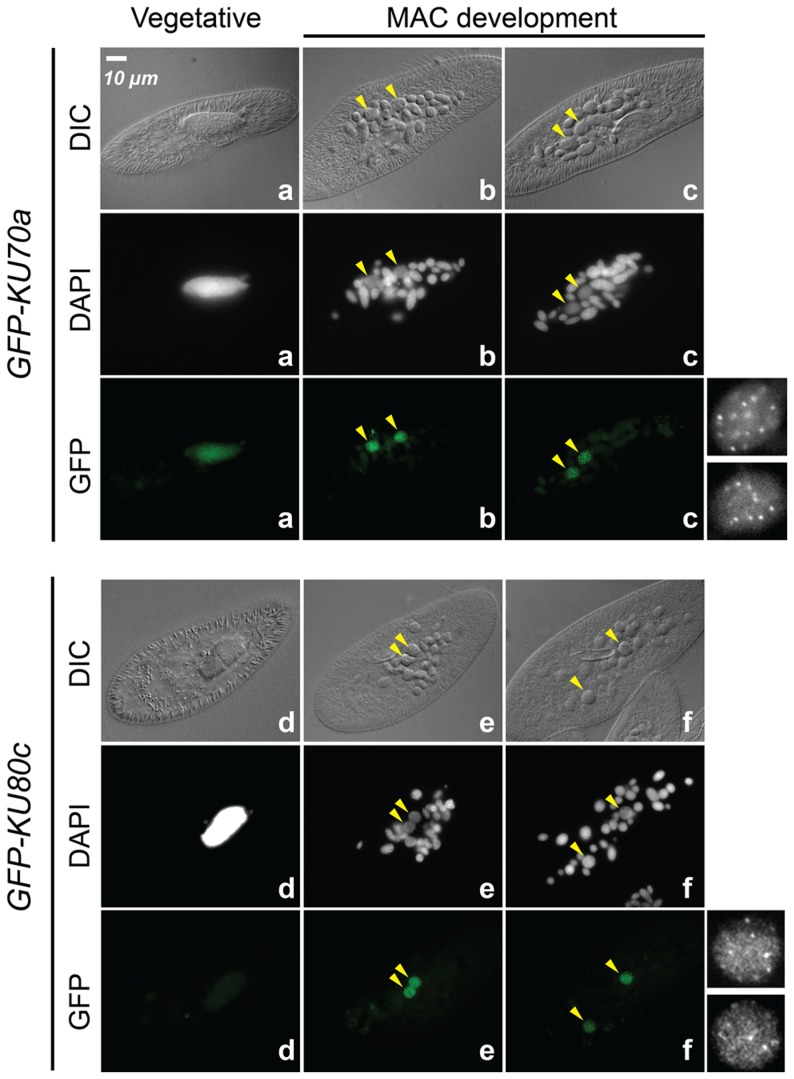
Figure 2. Nuclear localization of GFP-Ku fusions during autogamy.
Cells were microinjected with fusion transgenes expressing GFP-Ku70a (panels a, b, c) and GFP-Ku80c (panels d, e, f) under the control of their respective transcription signals. In autogamous cells, developing MACs are indicated by yellow arrowheads, the other DAPI-stained nuclei are fragments from the old vegetative MAC. For each protein, the GFP fluorescence sometimes concentrated in nuclear foci of unclear biological significance (see cells in panels c and f, and enlarged inserts on the right). In this particular experiment, expression of the GFP fusions had no significant effect on the recovery of viable post-autogamous progeny (87% progeny with functional new MACs for GFP-Ku70a, 90% for GFP-Ku80c).

