FIGURE 3:
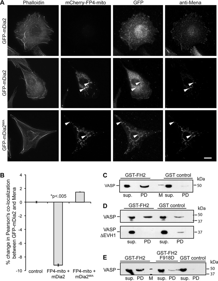
VASP directly interacts with mDia2. (A) Mitochondria-targeting assay. Transiently transfected NIH3T3 cells producing the indicated GFP- or mCherry-tagged proteins were chemically fixed and probed with a marker for F-actin (phalloidin) or antibody directed against Mena. Arrowheads indicate mitochondria labeled with mCherry-FP4-mito. Scale bar, 15 μm. (B) Quantification of A showing percentage change in Pearson's colocalization coefficient between Mena and GFP-mDia2. (C–E) Pull-down studies analyzing complex formation between mDia2 and VASP. Bead-immobilized GST-FH2 domain was incubated with purified VASP or VASPΔEVH1, and protein retained on beads was detected by SDS–PAGE and immunoblot. sup., supernatant; PD, pull down; M, molecular weight marker. (C) VASP interacts with the mDia2 FH2 domain. (D) VASP binds mDia2 through its EVH1 domain. (E) The FPPP motif in mDia2 FH2 mediates interaction with VASP. All data are from at least three independent experiments.

