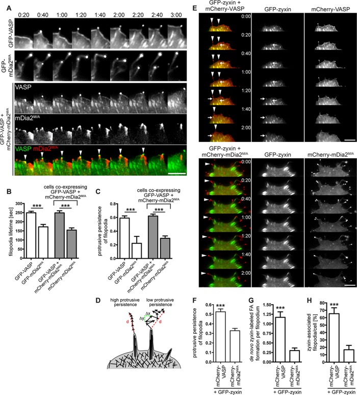
FIGURE 4:
VASP is essential for persistent protrusion of mDia2-generated filopodia. (A) VASP restores protrusive behavior of mDia2M/A-induced filopodia. MVD7 cells were transfected as indicated and plated on fibronectin-coated glass bottom dishes for 24 h before live-cell imaging. Montages of single frames from 3:00 min of live-cell microscopy time series. (B, C) Quantification of protrusive persistence (B) and lifetime (C) of filopodia from experiments shown in A. Filopodia tips in cells expressing GFP-VASP, GFP-mDia2M/A, or GFP-VASP plus mCherry-mDia2M/A were tracked in individual frames of 5-min movies, and so were filopodia that were in the same coexpressing cells but that contained only mCherry-mDia2M/A. Data represent at least two repetitions. n = 7–16 cells. (D) Distinct filopodia dynamics results in variable protrusive persistence. Protrusive persistence of filopodia was calculated by measuring the total distance (d) traveled by the filopodium tip divided by the sum of the change in directionality (Δx, Δy) of the filopodium tip between individual frames (see Materials and Methods). (E–H) VASP- but not mDia2M/A-containing filopodia (arrowheads) induce de novo assembly of GFP-zyxin–labeled focal adhesions at their base or in the shaft while actively protruding (arrows). MVD7 cells were transfected as indicated and plated on fibronectin-coated glass-bottom dishes for 24 h before live-cell imaging. (E) Montages of single frames from 2 min of live-cell microscopy time series. (F) GFP-zyxin expression does not change filopodia dynamics. (G) Quantification of de novo focal adhesion formation in protruding filopodia from experiments shown in E. (H) Percentage of zyxin-associated filopodia/cell. Data in F–H represent at least two repetitions. N = 8–11 cells, n = 41–60 filopodia in F and G; n = 58 (VASP) and 1426 (mDia2M/A) filopodia in H. ***p < 0.001. Scale bars, 5 μm.

