
FIGURE 5:
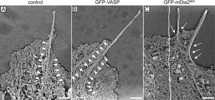
Actin bundles in filopodia initiated by mDia2M/A are not anchored in the lamellipodium actin network. Representative platinum-replica electron micrographs of actin cytoskeletons from MVD7 cells. Filopodium F-actin bundles from control cells (A) and cells complemented with GFP-VASP (B) are deeply embedded in the actin network of the lamellipodium (arrowheads) and splay apart at the filopodium root. In contrast, filopodia actin bundles of cells producing GFP-mDia2M/A (C) display poor connection to the underlying cortical cytoskeleton and do not have splayed roots (arrows), indicating that they may have formed via a different mechanism. Electron micrographs are representative images from at least three independent experiments. Scale bars, 0.5 μm.