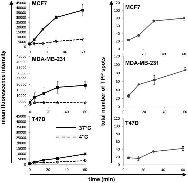
Figure 5. Kinetics of TPP internalization.
(Left panel) Flow cytometric analysis of memHsp70 positive human breast cancer lines MCF7, MDA-MB-231, T47D reveals an accumulation of the fluorescence intensity after incubation with CF-labeled TPP at 37°C between 1 and 60 min (solid lines). At 4°C (dashed lines) mean fluorescence intensity remained low within the same time frame. Cell lines with a high percentage of Hsp70 membrane positive cells (MCF7, MDA-MB-231) exhibit a higher and more rapid uptake of TPP, whereas the cell line with low Hsp70 membrane expression (T47D) exhibits only a low uptake of TPP. (Right panel) Confocal microscopy images were analyzed to provide a total count of fluorescent spots per cell after incubation with the TPP at 37°C for 30 min. Although fluorescent spots progressively accumulated in all three cell lines, this was most apparent in the MCF7 and MDA-MB-231 cell lines that express higher levels of memHsp70 than T47D cells.

