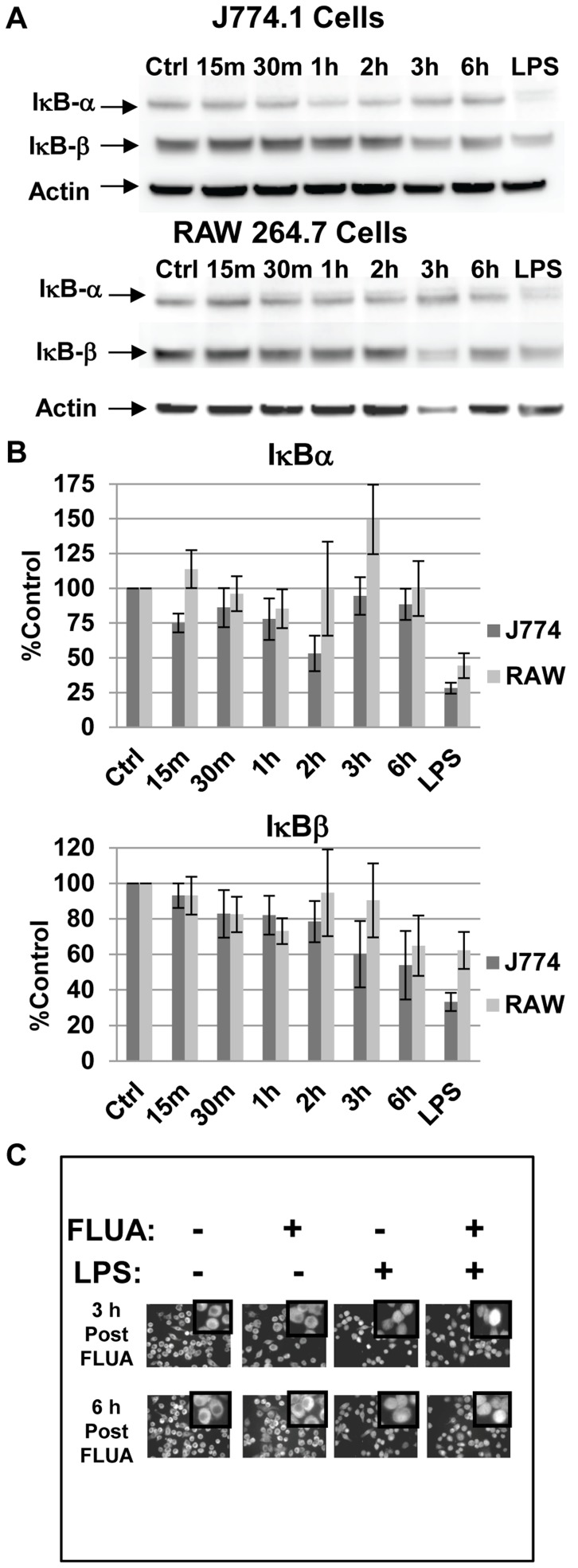
Figure 1. NFkB is not activated by FLUA at early time-points.
A. In J774.A1 cells IκBα levels were reduced at 2 h while IκBβ levels showed a gradual decline across the time points (but approximated control levels at 2 h). In RAW 264.7 cells, IκBα stayed near or above control levels while IκBβ levels showed a mild decline. Representative Western blots of cell lysates collected at the indicated time points post exposure to X-31 FLUA. Control and LPS treated (1 µg/ml for 15 minutes) cells were mock infected. B. Densitometry graphs from the Western blots. Data are normalized to actin, background is subtracted and protein levels are expressed as percentage of the mock infected control. Means are shown ± SEM, n = 3 to 6 independent experiments depending on time point. C. Nuclear localization of NFκB subunit p65 is not increased by X-31 infection of J774.A1 cells. Cells were infected or mock infected for the indicated periods, fixed, permeabilized and stained with anti-p65 antibody. LPS cells received 1 µg/ml LPS 30 minutes before fixation. LPS did increase NFκB nuclear localization as indicated by immunofluorescence. LPS activation was not blocked by prior X-31 exposure. 60X scale fields are shown with 255X digitally zoomed insets.

