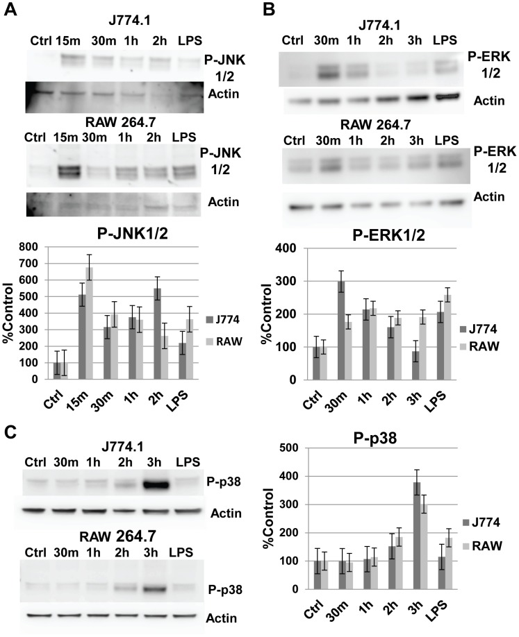
Figure 4. MAPKs are rapidly phosphorylated following FLUA infection.
A. J774.A1 and RAW 264.7 cells rapidly phosphorylate JNK 1/2 in response to X-31 exposure. In both cell lines maximal response was reached by 15 minutes post virus exposure. Control and LPS treated (1 µg/ml for 15 minutes) cells were mock infected. Representative Western blots and densitometry graphs from the Western blots (data are mean ± SEM, n = 5 independent experiments). B. J774.A1 and RAW 264.7 cells rapidly phosphorylate ERK 1/2 in response to X-31 exposure. In J774.A1 cells maximal response was reached by 30 minutes. RAW 264.7 cells had a lower maximal expression that was reached by 1 h. Control and LPS treated (1 µg/ml for 15 minutes) cells were mock infected. Representative Western blots and densitometry graphs from the Western blots (data are mean ± SEM, n = 4 independent experiments). C. J774.A1 and RAW 264.7 cells phosphorylate p38 in response to X-31 exposure. In both cell lines maximal response was reached by 3 h, a noticeable delay compared to the JNK1/2 and ERK1/2 activations. The antibody used recognizes α, β and γ p38 isoforms. Control and LPS treated (1 µg/ml for 15 minutes) cells were mock infected. Representative western blots and densitometry graphs from the western blots (data are mean ± SEM, n = 5 independent experiments).

