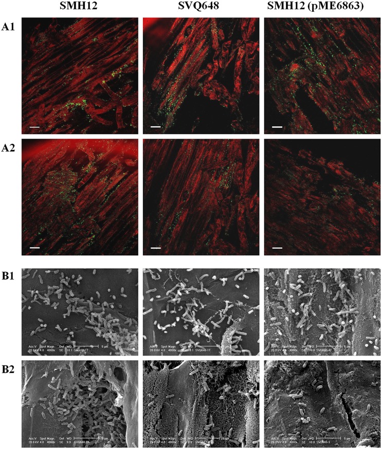
Figure 1. Visualization of the soybean root colonization.
A. Epifluorescence microscopy analysis of the colonization of the soybean rhizosphere by gfp-tagged bacteria [SMH12, SVQ648, SMH12 (pME6863)]. Roots were visualized 7 days after inoculation. 1. Proximal root. 2. Lateral roots. Bar, 100 µm. B. Scanning microscopy analysis of the colonization of the soybean rhizosphere by SMH12, SVQ648 and SMH12 (pME6863). Roots were visualized 7 days after inoculation. 1. Proximal root. 2. Lateral roots. Bar, 5 µm. SMH12: wild-type, SVQ648: nodD1 mutant, SMH12 (pME6863): lactonase strain.

