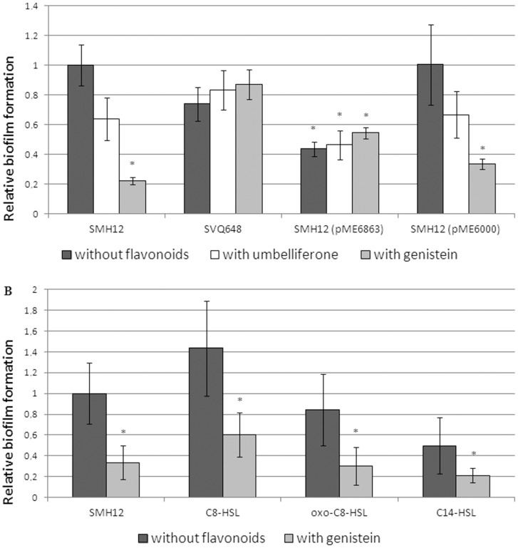
Figure 5. Adhesion of S. fredii SMH12 and derivatives on polystyrene surfaces.
Biofilms were measured as the amount of crystal violet absorbed by the biofilm formed on multi-well plates and determined by absorbance at 570 nm after de-staining with ethanol (see methods). Absorbance of the wild-type strain was arbitrarily given a value of 1. Averages and standard deviations of eight replicas per strain corresponding to five independent experiments are shown. The asterisks indicate a significant different at the level α = 5% with respect to wild-type strain by using the Mann-Whitney non-parametrical test. A. Dark gray bars correspond to experiments performed without flavonoids, white to experiments with umbelliferone and light grey bars to experiments with genistein. B. Dark gray bars correspond to experiments performed without flavonoids and light grey bars to experiments with genistein. 3-oxo-C8-HSL and C8-HSL are used at 5.5 µM. C14-HSL is used at 55 µM. SMH12: wild-type, SVQ648: nodD1 mutant, SMH12 (pME6863): lactonase strain. SMH12 (pME6000): carrying the empty plasmid.

