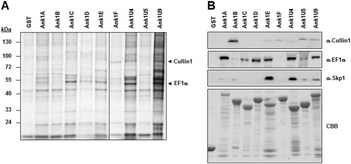
Figure 4. Identification of cellular proteins that interact with Ank proteins.
(A) Glutathione-Sepharose beads containing GST or one of the nine Ank proteins fused with GST were mixed with ECV304 cell lysate. Cellular interacting proteins were resolved by SDS-PAGE and visualized by Coomassie brilliant blue staining. Arrows indicate Cullin1 and EF1α, which were identified by mass spectrometry. (B) Immunoblot analyses were performed using specific antibodies and the cellular protein precipitates obtained from GST pull-down assays. At the bottom of the image, GST and the recombinant Ank proteins used in the pull-down assays are visualized after Coomassie brilliant blue (CBB).

