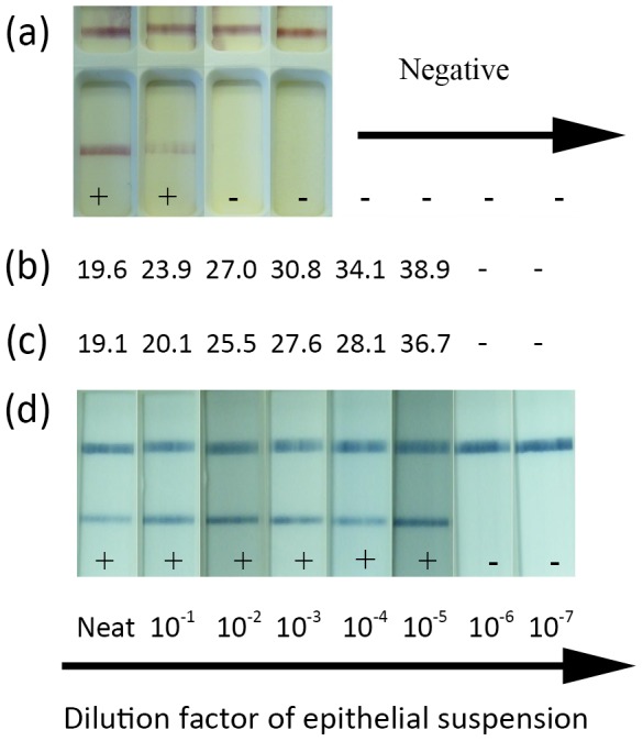
Figure 4. Comparative limit of detection between Svanova LFD, RT-qPCR, RT-LAMP-LFD and the RT-LAMP assays.

10 fold dilutions of FMDV containing epithelial suspensions were analysed using the Svanova LFD device (a) by applying the suspensions directly to the device. RNA was extracted from each suspension and subsequently analysed using either the RT-qPCR (giving a Ct value) (b) the RT-LAMP assay read using PicoGreen fluorescence(giving a Tp value) (c) or by mixing each 10 fold dilution 1∶5 with nuclease free water, and directly adding this to the RT-LAMP-LFD assay for subsequent detection with the LFD (d).
