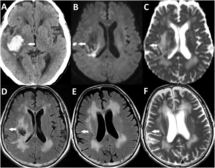
Figure 1. Example of a DWI lesion not visible on follow-up MRI.
(A) CT showing ICH in right putamen (arrow). (B, C and D) Baseline MRI showing a DWI lesion that has high signal intensity on DWI (B), low signal intensity on ADC (C) and equivocal high to intermediate signal intensity on FLAIR (D). (E and F) The DWI lesion is not visible on FLAIR (E) and T2WI (F) MRI 3 months after ICH.

