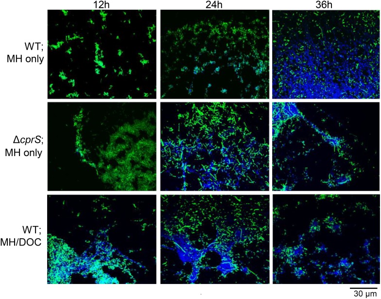
Figure 1. DNA appears in WT biofilms following attachment and is more pronounced under conditions that promote biofilm formation.
Biofilms of WT or ΔcprS were grown on glass coverslips in MH broth alone or MH/DOC (0.05%). At indicated times post-inoculation, coverslips were fixed, stained with DAPI, and visualized by confocal microscopy. Green: GFP-expressing bacteria; Blue: DAPI-stained DNA.

