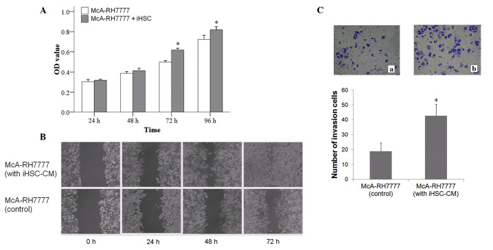
Figure 5.
(A) CCK-8 assay demonstrating iHSC-CM promotion of the proliferation of HCC cells (n=6 in each group; *P<0.05). (B) Scratch test demonstrating that CM from iHSCs increased the migration of HCC cells. (C) Transwell assay demonstrating that the number of migrated cells increased significantly when HCC cells were incubated with iHSC-CM and that iHSC-CM promoted the invasion of HCC cells. In the experimental group (McA-RH7777 + iHSC-CM) and the control group (McA-RH7777), the number of migrated cells was 42.5±7.9 and 18.8±5.5, respectively, demonstrating a significant difference (n=5 in each group; P<0.05). * P<0.05, compared with the control. Quantitative data are presented as the mean ± standard deviation. OD, optical density; CCK-8, cell counting kit-8; iHSCs, induction-activated HSCs; CM, conditioned medium; HCC, hepatocellular carcinoma.

